Time and access to imaging are everything when someone suffers a stroke. The time it takes for a patient to reach the hospital, be assessed in the emergency room, and then receive treatment is life-altering.
We screen everyone who comes to VGH with acute stroke symptoms using a CT scan of the brain and a CT angiogram of the head and neck. Without the CT scanner, we don’t know if a patient has a bleed in the brain, a tumour, or is suffering a stroke: the images give physicians a clear picture of the problem and provide vital information regarding potential treatment options.
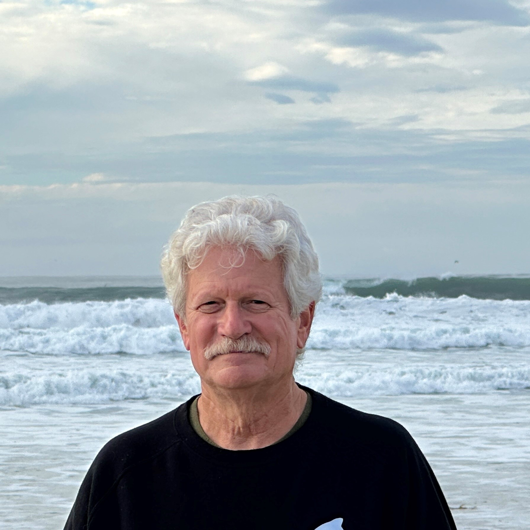
When I first saw Doug, he had what’s called an NIH Stroke Scale of 15, which is a disabling stroke deficit. He had lost the ability to speak and move the right side of his body.
Following the CT scan, a clot busting drug called tPA can be administered to patients who have suffered a stroke within a certain window of time (less than 4.5 hours since last being seen normal). Doug was a perfect candidate for tPA. The emergency CT scan of his brain confirmed a large blockage of a major artery in his brain and revealed that there was a large volume of brain tissue that would die if the artery did not soon open.
The procedure we use to remove a clot like Doug’s is called an emergency embolectomy for an intracranial occlusion. It involves accessing an artery through the groin and guiding a special device through the aorta into the major artery in the brain to access the blockage. A stent or suction device then physically pulls the clot out of the brain, restoring blood flow to the brain and reducing brain damage related to stroke. In most cases we save a large amount of brain from irreversible injury, and in some cases the stroke symptoms may disappear almost immediately.
In Doug’s case, everything worked out perfectly. Although the procedure can take an hour or more, in Doug’s case normal blood flow to the brain was achieved within half an hour. Ten minutes after the procedure, Doug had no clinical stroke deficits and two days later, he was discharged and walked out the front door of the hospital.
A perfect outcome with Doug’s type of stroke is rare; less than five percent of patients in his situation might have zero stroke deficits long term. But great outcomes—life-changing ones—happen every day at VGH. The CT Scanner plays a vital role in this.
I take a moment to thank the supporters of the Victoria Hospitals Foundation for helping to upgrade one of our CT scanners; it is desperately needed. In my scope of work with stroke patients, we simply could not treat people without CT imaging. Through your donation, you are enabling care, and truly, you are helping save lives.
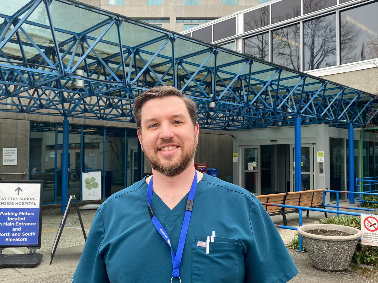
—Dr. Andrew Lockey
Stroke Neurologist, Victoria General Hospital


