Our radiology team remembers Mary-Gayle well. We do rounds every week, and would discuss her case in great detail due to the sheer amount of medical imaging that was involved in her complex care journey.
Over the course of Mary-Gayle’s treatment, we used the entire spectrum of medical imaging equipment—including CT, C-Arm, and MRI technology.
With aortic aneurysms like Mary-Gayle’s, we use CT scanners to measure the ballooning aneurysm sac over time. This monitoring allows us to determine if and when surgery is required. Aortic aneurysms are considered at low risk of bursting until the sac reaches five centimeters in size. At this point, the first option is to try inserting a stent. This is a small metal mesh and fabric tube which serves as re-enforcement for the weak area of blood vessel that is ballooning. With guidance from X-Ray and C-Arm imaging, this tube is inserted through one of the patient’s arteries and set in place.
Unfortunately, one of the challenges that can come with this procedure is that if there is any kind of leakage past the stent into the existing sac, the stent restricts the exit pathway for that blood flow. This can cause the sac to expand more rapidly and eventually burst—and in Mary-Gayle’s case, it did. This is when interventional radiologists come in to find an alternative treatment.
In my line of work, I commonly use a medical glue known as Onyx. It’s a substance that bonds instantly when exposed to blood. Our plan with Mary-Gayle was to plug up the sac with Onyx to seal any leaks and prevent the sac from filling with any more fluid in the future. This requires great precision.
In order to identify the best injection spot, we utilized a recent innovation in MRI technology known as 4D MRI. This new technique allows us to make multiple scans per minute, giving us a dynamic picture of how blood is flowing. From there, we can identify exactly where the leaks are and where we should inject. However, many readers will know that due to an MRI machine’s powerful magnets, metal objects—including needles—cannot be taken into the MRI room. So, to actually make the injection, we moved to the CT room where our MRI images could be overlaid with CT scans to guide our needle placement. The procedure was precise and successful. One more scan in the MRI confirmed that the leak had completely stopped.
Mary-Gayle’s journey is a prime example of the importance of medical imaging technology. Just like any other industry, our hospitals rely on investments in technology to advance the work we do. Without our donors’ help, we wouldn’t have the leading-edge medical imaging equipment needed to treat patients like Mary-Gayle. Through your generosity, we can create more success stories like hers!
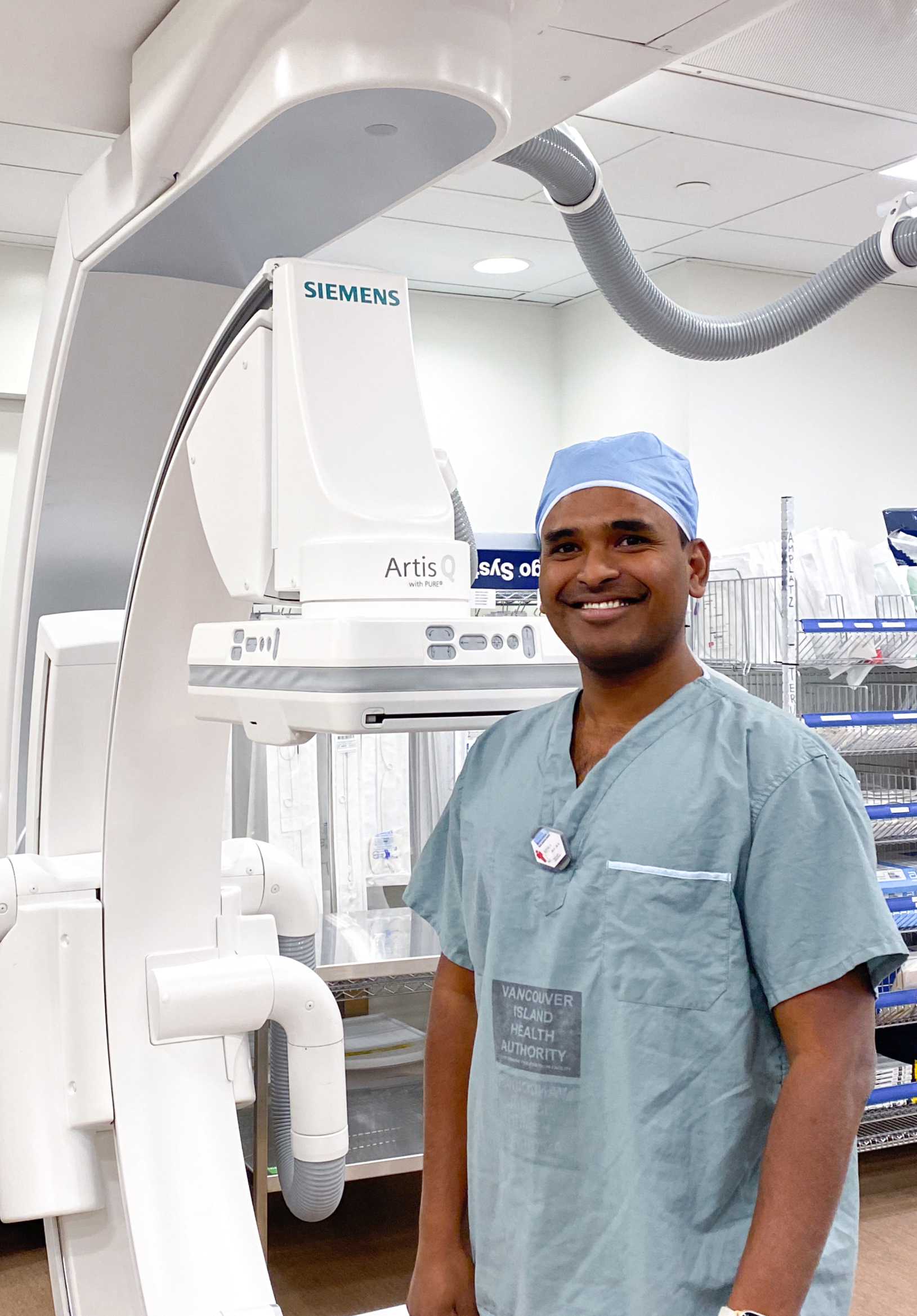
— Dr. Vamshi Kotha
Interventional Radiologist, Royal Jubilee & Victoria General hospitals


